Age-Related Macular Degeneration (ARMD)
Introduction
Age-Related Macular Degeneration (ARMD) is the leading cause of vision loss in older American adults. Patients are referred by their general ophthalmologist or optometrist to retina specialist for further evaluation, treatment and care. Patients afflicted with macular degeneration may note a worsening of their central vision while retaining good peripheral vision.
Many patients with macular degeneration share a common misconception and fear that the disease will automatically progress to blindness. Macular degeneration only affects the central field of vision. The macula is the small central part of the retina responsible for seeing fine details. The macula is assist patients perform activities such as reading and driving. In macular degeneration, patients suffer varying degrees of destruction to this tissue and can experience a wide spectrum of visual problems. These include, but are not limited to, blurred central vision, increasing difficulty with reading, patchy visual loss, and the distortion or warping of straight objects. However, even patients with advanced macular disease tend to retain excellent peripheral vision.
What causes macular degeneration and who is at risk?
Many older people develop macular degeneration as part of the body’s natural aging process, although cases have been reported in younger patients. Exactly why it develops is not known, and no treatment has been uniformly effective. Macular degeneration is a leading cause of severe vision loss in patients over 65. Additionally, a number of risk factors predispose certain people to the development and progression of the disease, including:
Cigarette smoking
Having a family member with macular degeneration.
Regarding the last risk factor, much research has been done regarding the genetics of macular degeneration. Although there is currently no way to definitively predict who will or will not develop macular degeneration, those with affected relatives are at significantly higher risk. As a result, adult relatives of macular degeneration patients should schedule an appointment with a retinal specialist and undergo a baseline evaluation and dilated exam.
Are there different types of Macular Degeneration?
Generally, patients with macular degeneration are grouped into one of two types– Dry Age Related Macular Degenerationthose with dry macular degeneration and those with wet macular degeneration.
Dry Macular Degeneration (Atrophic)

Most people have the “dry”: form of degeneration. It is caused by aging and thinning of the tissues of the macula. Usually vision loss is gradual.
Wet Macular Degeneration (Exudative)
The “wet” form of macular degeneration accounts for about 10 percent of all cases. These cases develop significant vision loss. Wet degeneration result when Wet Macular Degenerationabnormal blood vessels from underneath the retina at the back of the eye grow and leak. These new blood vessels leak fluid or blood causing the vision to blur. Vision loss is sudden and severe.
What are the symptoms of macular degeneration? Visual symptoms can differ from person to person. The condition can be hardly noticeable in its early stages. Sometimes only one eye loses vision while the other eye continues to see well. When both eyes are affected, the loss of central vision may be notices much more quickly.
Some common visual symptoms are:
- a dark or empty area appears in the center of vision;
- straight lines look distorted;
- words on a page look blurred;
- What Tests are Helpful in Aiding Diagnosis?
Sometimes it is difficult to differentiate between dry and wet macular degeneration, so our physicians will use a variety of clinical tests and exam techniques to make an accurate diagnosis.
During an initial visit for macular degeneration, patients typically undergo several different test:
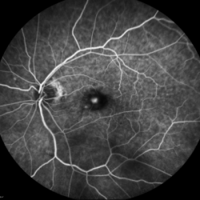
Fluorescein Angiography (FA), ICG and Optical Coherence Tomography (OCT). FA is for differentiating between dry and wet ARMD. It involves the injection of a small amount of vegetable-based dye through a patient’s vein, usually the arm or hand. A technician will take a series of retinal photographs. The injected dye lights up the retina’s vascular network and helps our physicians focus in on the area of leakage in those patients with wet macular degeneration. This test is useful in determining the extent and progression of the disease, and also helps when targeting specific treatment for macular degeneration.
OCT is a non-invasive, quick exam that is now used by many ophthalmologists. Using reflected light rays, OCT provides a detailed, highly magnified, cross-section view of a patient’s macula. Sometimes, it will uncover tiny areas of leakage not readily apparent to a retina specialist during a microscopic exam.
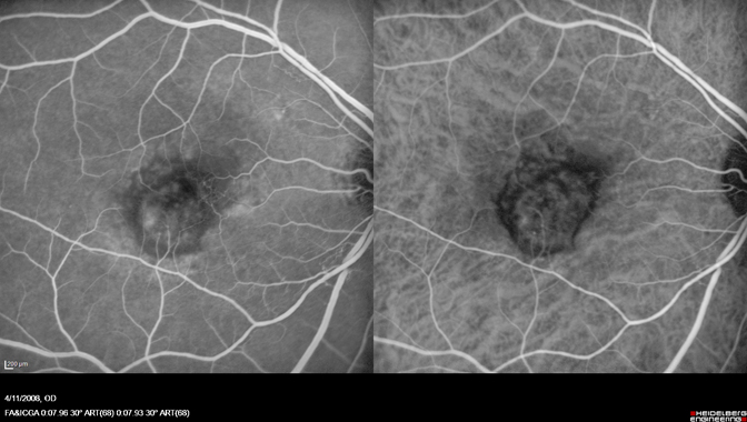
Wet ARMD
Patients Play an Important Role in Diagnosis and Care
Even though the advances in ophthalmic imaging help our doctors detect early changes, there is no single test that can be relied upon to direct treatment and clinical decision-making. A patient’s relationship with their retina specialist is at the center of our good patient-physician communication.. When making important decisions about retinal health, it is very much a joint effort. The patient’s perception of his or her vision changes, however slight, is every bit as important in directing care as are the clinical tests.
As a result, every patient diagnosed with macular degeneration should establish a daily routine for monitoring their own vision. This is most simply and reliably performed with an Amsler grid, a simple graph paper-like patchwork of straight vertical and horizontal lines, which we are happy to provide at no cost – simply ask one of our technicians.
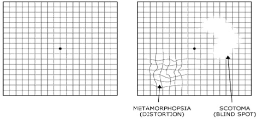
Amsler GridOften, patients with subtle progression of macular disease will report new “waviness” or “missing areas” when monitoring their grid. Should you or a family member notice new changes on an Amsler grid, contact us promptly. One of our retina specialists or trained ophthalmic nurses is always available to consult with you by phone. As a general rule, patients who receive treatment in a more timely fashion tend to fare better than those who delay evaluation by a retinal specialist.
What Treatments are Available?
Dramatic progress has been made in the effort to treat macular degeneration related vision loss. The most promising new treatments are anti VEGF drugs which help block certain growth factors that are thought to cause “leaky” blood vessel growth in wet macular degeneration. These medications are introduced directly into the eye through a tiny needle. While the thought of an “eye shot” might cause many to feel anxious, there is little, if any, patient discomfort, and the possible visual gains far outweigh any fears.